I asked Hiroki Yokota the other day about LIPUS and he mentioned something interesting: "Ultrasound is considered as high frequency vibration, and I believe that most people think this as a unique form of mechanical stimulation." If Ultrasound is considered high frequency vibration which induces fluid flow, what if there are other cheaper ways of inducing vibration(a LIPUS machine can run you $3,000, there are cheaper ones for about $60-100 but their effectiveness has not been as well studied: Porable Home Ultrasound Unit
Mechanical signal influence on mesenchymal stem cell fate is enhanced by incorporation of refractory periods into the loading regimen.
"Mechanical signals of both low and high intensity are inhibitory to fat and anabolic to bone in vivo, and have been shown to directly affect mesenchymal stem cell pools from which fat and bone precursors emerge[chondrocytes are bone cell precursors]. To identify an idealized mechanical regimen which can regulate MSC fate, low intensity vibration (LIV; <10 microstrain, 90Hz) and high magnitude strain (HMS; 20,000 microstrain, 0.17Hz)[20,000 microstrain is crazy as according to the mechanostat model 15,000 microstrain is the point of fracture] were examined in MSC undergoing adipogenesis. Two×twenty minute bouts of either LIV or HMS suppressed adipogenesis when there was at least a 1h refractory period between bouts; this effect was enhanced when the rest period was extended to 3h. Mechanical efficacy to inhibit adipogenesis increased with additional loading bouts if a refractory period was incorporated. Mechanical suppression of adipogenesis with LIV involved inhibition of GSK3β[Remember Lithium inhibits GSK3Beta] with subsequent activation of β-catenin as has been shown for HMS. These data indicate that mechanical biasing of MSC lineage selection is more dependent on event scheduling than on load magnitude or duration. As such, a full day of rest should not be required to "reset" the mechanical responsiveness of MSCs, and suggests that incorporating several brief mechanical challenges within a 24h period may improve salutary endpoints in vivo[maybe loading once every 24 hours isn't the best for LSJL?]. "
The study found that mechanical loading increased sensitivity of stem cells to BMP2 but the response was osteogenic. The GSK3Beta inhibition may be important so as to activate Nfatc1 and Cox2.
Low Intensity Vibration inhibited GSK-3Beta which can increase height in growth plate chondrocytes but this study doesn't indicate that LIV increased chondrogenic markers like TGF-Beta[bone cells(which differentiation is encouraged by the activation of Beta Catenin) can increase height but not as effectively as chondrocytes]. But LIV produces very similar effects to High levels of Mechanical Loading[20,000 microstrain which is past the point of fracture] so LIV is superior to ordinary(non-lateral) mechanical loading.
This study indicates that Ultrasound may have a beneficial affect other than shear strain alone as was previously believed:
Low-Intensity Pulsed Ultrasound Modulates Shear Stress Induced PGHS-2 Expression and PGE(2) Synthesis in MLO-Y4 Osteocyte-Like Cells.
"Fluid shear stress (SS) has been shown to be a prevailing physiological stimulus in the regulation of bone cell metabolism and so are the exogenous biomechanical forces, like ultrasound (US) and vibration. The purpose of this study is to elaborate the interplay of laminar fluid SS with low-intensity pulsed US in the regulation of prostaglandin H synthase 2 (PGHS-2)[precursor to PGE2] and prostaglandin E(2) (PGE(2))[involved in bone turnover]. Murine long bone osteocyte-like (MLO-Y4) cells were exposed to various regimes of US (1.5 Hz, 30 mW/cm(2)) and SS (19 dyn/cm(2)) alone and sequentially. Changes in PGHS-2 gene expression levels were quantified at 3 and 24 h using real-time RT-PCR. PGE(2) levels in the culture media were measured using enzyme immunoassay at 3 and 24 h. PGE(2) levels significantly increased after exposure to SS for 3 and 24 h by 2.17 +/- 0.02 and 5.47 +/- 0.42-fold, respectively, compared to control cells. A 20 min US treatment prior to SS significantly increased SS PGE(2) levels 2.95 +/- 0.18 and 2.90 +/- 0.50-fold at 3 and 24 h, respectively. US also significantly increased PGHS-2 mRNA levels in cells exposed to SS. SS caused a 2.74 +/- 0.49-fold increase in PGHS-2 mRNA levels at 3 h and a significant 3.70 +/- 0.25-fold increase at 24 h relative to control. A 20 min US treatment caused 1.35 +/- 0.49 and 2.44 +/- 0.82-fold increase in PGHS-2 mRNA levels in cells exposed to SS at 3 and 24 h, respectively. These results indicate that combining US with SS may have a more anabolic benefit for bone tissue than either stimulus alone."
PGE2 isn't something likely to increase height although osteoclasts due play a role in height growth. This study does show that Ultrasound may have an effect other than fluid flow shear stress(and in turn Vibration and LSJL) on bone cells(and in turn possibly stem cells and chondrocytes).
Pressure differs from Shear Strain as shown by this study(vibration & Ultrasound are only likely to induce shear strain & not pressure, LSJL induces both):
A new bioreactor for the controlled application of complex mechanical stimuli for cartilage tissue engineering.
"A new bioreactor is described where different mechanical stimuli, i.e. shear stress and hydrostatic pressure, can be combined in different ways to study the mechanobiology of tissue engineered cartilage. Shear stress is imposed on cells by forcing the culture medium through the scaffolds, whereas a high hydrostatic pressure up to 15 MPa is generated by pressurizing the culture medium. Fluid-dynamic experimental tests have been performed and successful validation of the bioreactor has been carried out by dynamic culture of tissue-engineered cartilage constructs."
Remember it was hydrostatic pressure that resulted in the organic development of chondrocytes.
Mechanical vibrations increase the proliferation of articular chondrocytes in high-density culture.
"Mechanical vibrations can enhance chondrocyte proliferation in monolayer culture. Thus, it was hypothesized that chondrocytes grown in high-density culture would respond in a similar fashion while maintaining phenotypic stability. Isolated bovine articular chondrocytes were seeded in high-density culture on Millicell filters and subjected to mechanical vibrations 48 h after seeding. Mechanical vibrations enhanced chondrocyte proliferation at frequencies above 350 Hz, with the peak response occurring at a 1g amplitude for a duration of 30 min. Under these conditions, the gene expression of cartilage-specific and dedifferentiation markers (collagen II, collagen I, and aggrecan) were unchanged by the imposed stimulus. To determine the effect of accumulated extracellular matrix (ECM) on this proliferative response, selected cultures were stimulated under the same conditions after varying lengths of preculture. The amount of accumulated ECM (collagen and proteoglycans) decreased this proliferative response, with the cultures becoming insensitive to the stimulus after 1 week of preculture. Thus, mechanical vibration can serve as an effective means preferentially to stimulate the proliferation of chondrocytes during culture, but its effects appear to be limited to the early stages where ECM accumulation is at a minimum."
This can also be involved in the increasing load needed to get a response from LSJL. ECM can accumulate over time making it harder and harder to induce stimulus.
Hydrostatic pressure unlike low frequency vibration has been shown to induce stem cell proliferation into chondrocytes. Therefore, LIPUS may not be an alternative to LSJL but rather a way to augment LSJL by increasing chondrocyte proliferation. It may also be a way to grow taller during growth plate development by inducing vibration on the growth plate chondrocytes and increasing cellular proliferation there.
Effects of vibration and hyaluronic acid on activation of three-dimensional cultured chondrocytes.
"Chondrocytes were obtained from metatarsophalangeal joints of freshly killed 6-month-old pigs. Twenty-four-well plates containing type I collagen sponge disks were used to culture samples. The frequency and the amplitude of the vibration of the well plate were 100 Hz and 0.5 nm, respectively. We produced 3-dimensional cartilage tissue using HA and vibration with collagen sponge as a carrier. Four different culture conditions were examined: a control HA-Vib- group, an HA-Vib+ group, an HA+Vib- group, and an HA+Vib+ group. Each group was cultured for 2 weeks. After culture days 3, 7, 10, and 14 (every 3.5 days), the levels of chondroitin 4-sulfate (C4S) and chondroitin 6-sulfate (C6S) isomers synthesized in each culture medium were measured. Histologic analysis, immunohistochemical analysis, and electron microscopic examination were performed.
Mean C4S and C6S synthesis had increased rapidly after 7 days of culture and continued to increase thereafter. There were significant differences among the 4 groups (P < 0.01). Synthesis of both C4S and C6S was most abundant in the HA+Vib+ group and the lowest in the HA-Vib- group[almost 50% greater than the control group]. After 1 and 2 weeks of culture, the chondrocytes had formed stratified structures on the collagen sponges in all groups, although the thickest structure was observed in the HA+Vib+ group and the thinnest in the HA-Vib- group. Under immunofluorescence, the HA+Vib+ group exhibited the strongest chromatic features. Under electron microscopy, the chondrocytes in the HA+Vib+ group exhibited many long and slender prominences on their surface, and extracellular substance could be observed associated with the cells.
Our results indicate that the combination of vibration and HA activates the production of proteoglycan in 3-dimensional cultured chondrocytes and stimulates MAPK and beta-catenin. This suggests that some mechanoreceptors for vibration exist on the plasma membrane of chondrocytes and activate the intracellular signal transduction system."
So Hyaluronic Acid and Vibration are both pro-chondrogenic.
"[Regarding] the effect of vibration on monolayer cultured chondrocytes and reported that the frequency of 300 Hz was most effective for promoting proteoglycan synthesis, whereas frequencies >400 Hz suppressed biosynthesis"<-Of couse this may be different for MSCs. This is 300 times per second much greater than the 5 times per second in the LSJL limb lengthening studies.
"The expression of proteins involved in the signal transduction system was significantly higher in the groups treated with vibration, strongly suggesting the existence of receptors for vibration on the plasma membrane of chondrocytes."
"One of the roles of CD44 is to transfer HA into the cell to be processed by hyaluronidase. RHAMM is known to regulate cellular motility and signal transduction in the cell cycle. Cell-surface RHAMM interacts with both HA and fibronectin to regulate phosphorylation of functionally important proteins which bind to actin and integrin."
"In order for chondrocytes to survive and multiply, close contact with the extracellular matrix must be maintained[and stem cells must get close together to form growth plates and HA provides a good base for mesenchymal condensation]. Integrin is involved in cellular adhesion by binding to a membrane-penetrating ligand to induce various biochemical changes inside cells. Adhesive stimuli, via integrin, elevate the kinase activity of FAK, and via Ras, the activity of MAPK is ultimately increased. MAPK is responsible for the conversion of many extracellular stimuli into specific cellular responses regulating proliferation, migration, differentiation, and apoptosis. MAPK pathways coordinate regulation of gene transcription and protein synthesis for adaptation to changes in the extracellular environment. In desmosomes, skeletal proteins (such as integrin, talin, vinculin, and paxillin) gather to form actin fibers. Phalloidin is a specific marker for actin fibers, and in the present study, phalloidin staining revealed the development of actin fibers in chondrocytes treated with HA and/or vibration. This suggests that vibration produces micro motion in the extracellular matrix causing actin fibers to be activated, thus allowing chondrocytes to strongly adhere to the extracellular matrix."
"Exogenous HA regulates the distribution of proteoglycan synthesized by chondrocytes, fills the gaps among matrices, and allows chondrocytes to more easily adhere to the extracellular matrix"<- you can get exogenous HA by taking HA supplements.
"We propose that vibration plays 3 important roles, as follows: first, vibration acts on the mechanoreceptors on the plasma membrane to activate the intracellular signal transduction system; second, vibration activates integrin-mediated adhesion to extracellular proteins; finally, vibration allows HA to be distributed evenly among chondrocytes and the extracellular matrix to move, thus improving delivery of nutrients to chondrocytes in deeper layers and transportation of waste products"
The response of vocal fold fibroblasts and mesenchymal stromal cells to vibration.
"The purpose of this study was to compare the response of human vocal fold fibroblasts (hVFF) and bone marrow mesenchymal stem cells (BM-MSC) to vibratory stimulus. In order to study these effects, a bioreactor capable of vibrating two cell seeded substrates was developed. The cell seeded substrates contact each other as a result of the sinusoidal frequency, producing a motion similar to the movement of true vocal folds. Utilizing this bioreactor, hVFF and BM-MSC were subjected to 200 Hz vibration and 20% strain for 8 hours."
"HVFF significantly proliferated (p = 0.011) when subjected to 200 Hz vibration and 20% strain, while BM-MSC did not (p = 1.0). A statistically significant increase in apoptosis of BM-MSC (p = 0.0402) was observed under the experimental conditions; however high cell viability (96%) was maintained."
"no significant differences in expression levels of collagen I (BM-MSC p = 0.1951, hVFF p = v0.3629), fibronectin (BM-MSC p = 0.1951, hVFF p = 0.2513), and TGF-β1 (BM-MSC p = 0.2534, hVFF p = 0.6029) between vibratory and static conditions in either cell type."
"ligament fibroblasts secrete collagen I and III when subjected to axial strain"
So vibration may not induce chondrogenic differentiation on it's own with this particular modality. However vibration of the whole bone may induce fluid flow which could induce chondrogenic differentiation.
Osteogenic differentiation of bone marrow-derived mesenchymal stromal cells on bone-derived scaffolds: effect of microvibration and role of ERK1/2 activation. states that vibration of MSCs on scaffolds which can involve fluid flow induces ERK1/2 phosphorylation. In contrast to the other study type I collagen was increased.
Unfortunately, there have been no studies of vibration inducing chondrogenic gene expression or chondrogenic differentiation.
Low-level mechanical vibrations can influence bone resorption and bone formation in the growing skeleton.
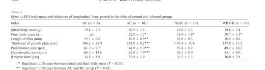
"Eight-week-old female BALB/cByJ mice were divided into four groups, baseline control (n = 8), age-matched control (n = 10), whole-body vibration (WBV) at 45 Hz (0.3 g) for 15 min day(-1) (n = 10), and WBV that were interrupted every second by 10 of rest (WBV-R, n = 10). In vivo strain gaging of two additional mice indicated that the mechanical signal induced strain oscillations of approximately 10 microstrain on the periosteal surface of the proximal tibia. After 3 weeks of WBV, applied for 15 min each day, osteoclastic activity in the trabecular metaphysis and epiphysis of the tibia was 33% and 31% lower than in age-matched controls. Bone formation rates (BFR.BS(-1)) on the endocortical surface of the metaphysis were 30% greater in WBV than in age-matched control mice but trabecular and middiaphyseal BFR were not significantly altered. The insertion of rest periods (WBV-R) failed to potentiate the cellular effects."
"After the 3-week experimental duration, the right tibia was harvested"
Low frequency vibration did not alter growth plate size nor longitudinal growth. Although there was a non-significant increase in growth plate thickness, proliferative zone, hypertrophic zone and decrease in reserve zone between whole body vibration groups and age maged controls. And in that case it was found that the bone length was about the same so in all likelihood there is more growth to be had. Growth plate parameters increased but bone length stayed the same therefore it is likely that the vibrated bone had more growth.

"Standing on an inactive plate induced strains on the order of 1 με. The vibratory oscillations applied at a frequency of 45 Hz and peak accelerations of 0.3 g induced peak bone strain oscillations at the antero-medial surface of the tibia on the order of 10 με."
The strain is the same as LSJL but maybe the frequency was too high to induce length gain or LSJL induces other effects besides microstrain.
Low-Level Accelerations Applied in the Absence of Weight Bearing Can Enhance Trabecular Bone Formation
"A loading apparatus, developed to shake specific segments of the murine skeleton without the direct application of deformations to the tissue, was used to subject the left tibia of eight anesthesized adult female C57BL/6J mice to small (0.3 g or 0.6 g) 45Hz sinusoidal accelerations for 10 min/day, while the right tibia served as an internal control."
"At 0.3 g peak accelerations, peak-to-peak strains induced in the proximal tibia were 1.1 microepsilons. Doubling peak acceleration to 0.6 g doubled the strains (2.2 me)."
"No significant differences in bone length were detected between accelerated and control tibiae"
No comments:
Post a Comment